Day 2 :
Keynote Forum
Shaofeng Yan
Dartmouth Hitchcock Medical Center, USA
Keynote: Application of molecular tools in diagnosis and treatment of cutaneous melanoma
Time : 09:30-10:10
Biography:
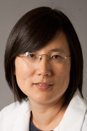
Shaofeng Yan has received her MD degree from Peking Union Medical College and PhD degree from University of Washington at Seattle. She has completed Anatomic and Clinical Pathology Residency at Dartmouth Hitchcock Medical Center and a combined Harvard Dermatopathology Fellowship at Massachusetts General Hospital, Brigham Women’s Hospital and Beth Israel Deaconess Hospital. She is currently the Director of Dermatopathology Section and Program Director of Dermatolopathology Fellowship Program of the Department of Pathology at Dartmouth Hitchcock Medical Center.
Abstract:
Melanoma is one of the most aggressive types of skin cancer. Histopathological criteria sometimes may be inadequate in differentiating difficult melanocytic lesions. Treatment for advanced melanoma cases still remains elusive. New development in molecular testing technologies is useful for improving diagnosis. New therapeutic approaches based upon a growing understanding of the underlying molecular abnormalities have been used in advanced malignant melanoma recently. Single nucleotide polymorphism (SNP) microarray analysis can accurately detect copy number changes and aid in improving differentiation of malignant melanoma from benign melanocytic proliferation. Next-generation sequencing (NGS) technologies have enabled detection of key genetic mutations for targeted therapy. Here we share our experience of application of SNP microarray analysis in differentiation of malignant melanoma from benign melanocytic proliferation. Furthermore using NGS testing for a 50 gene panel, we have identified numerous variable mutations, which may represent potential targets for future therapies in patients with advanced melanoma.
Keynote Forum
Vikash Sewram,
African Cancer Institute, South Africa
Keynote: The burden of melanoma in the Western Cape province of South Africa: Provision of a platform for evidence-based research
Time : 10:10-10:50

Biography:
Professor Vikash Sewram is the Chairperson of the Ministerial Advisory Committee on the Prevention and Control of Cancer in South Africa, the Founding Director of the African Cancer Institute and Professor of Community Health at the Faculty of Medicine and Health Sciences, Stellenbosch University, South Africa. He obtained a PhD degree in Medicinal Chemistry and Physiology from the University of Natal in 1998, an MPH in Cancer Epidemiology (with distinction) from the School of Public Health and Family Medicine, University of Cape Town, in 2002, and a PhD in Public Health: Cancer Epidemiology from the same university in 2007. In 2009 he was nominated to the Academy of Science of South Africa and, in 2014, to the Permanent Scientific Committee in the Oncology Section of the World Organization for Specialized Studies on Diseases of the Esophagus. He has spent time abroad as visiting scientist at the International Agency for Research on Cancer in Lyon, France; School of Public Health, University of Michigan, USA; and the Cancer Council NSW in Sydney, Australia. His research achievements have earned him 10 national and nine international research awards, and have resulted in numerous national and international collaborations, peer-reviewed publications, research grants and postgraduate student supervision.
Abstract:
Age standardised incidence rates of 15.88/100 000 and 12.68/100 000 for melanoma in South Africa have been reported nationally for Caucasian men and women respectively. The incidence rates in the Cape Town region of the Western Cape Province however are in excess of 60/100 000. Cutaneous melanoma (CM) has the highest incidence in Caucasians, followed by the persons of mixed ancestry and a considerably lower incidence in both the Black and Asian population. Over the years, the rates of melanoma has been increasing and to further study this disease the African Cancer Institute (ACI) at Stellenbosch University has embarked on the establishment of a melanoma research platform that encompasses primary prevention, behavioural sciences, genomic research, and public policy. Despite numerous treatment options becoming available, drug access remains a limiting step and melanoma prevention and control remain a core focus through the extensive network of partners within the public and private sector organizations.
Research within the Division of Dermatology has commenced on the molecular biology of hand and foot melanoma, also known as acral melanoma (AM), which appears to be a clinically distinct variant of melanoma. This variant of melanoma represents the most common expression of melanoma in the Black population, and has a 5-year survival rate of 80.3%, lower than for other forms of melanoma. This is thought to be a result of delays in diagnosis. AM is also thought to have unique patterns of genetic mutation, when compared to other forms of CM. The current studies aim to identify molecular alterations that drive tumorigenesis in AM in Southern Africans. It is anticipated that this study will help classify high from low risk AMs followed by the development of a molecular predictive test and to characterize the clinical and histological features of AM in our population.
By harnessing the collaborative intellect of individuals, groups and institutions throughout the region and abroad, the ACI seeks to strengthen and accelerate the translation of melanoma control knowledge into public health action.
Keynote Forum
Yohei Tanaka
Reconstructive Surgery and Anti-aging Center, Japan
Keynote: The necessity of solar near-infrared protection
Time : 11:10-11:50

Biography:
Yohei Tanaka is one of the leading Plastic Surgeons in Japan. He directs his clinic, Society for Near-infrared Rays Research and International Photobiological Society. He conducts many researches as a Visiting Professor of Niigata University of Pharmacy and Applied Life Sciences and Lecturer of Tokyo Women’s Medical University. He has published over 20 peer-reviewed papers in English and has edited 2 international open access books regarding near-infrared. His goal is to discover the most effective near-infrared wavelengths for rejuvenation and anti-cancer therapy and to further study solar near-infrared and how best we can protect ourselves against its photoaging.
Abstract:
Over half of the solar energy consists of near-infrared and intensive or long-term solar near-infrared exposure induces photoaging. Despite the wide prevalence of a variety of ultraviolet blocking materials, such as sunscreen, sunglasses, glasses, films, umbrellas and fibers that are useful in protecting the skin against ultraviolet exposure, solar near-infrared cannot be blocked and the necessity to protect against solar near-infrared has not been well recognized. Solar near-infrared can penetrate the skin and the sclera and affect the deeper tissues, including muscles, lens and retina with its high permeability. I have elucidated that solar near-infrared can induce various biological effects. Continual long-term exposure to solar near-infrared performs as an aging factor. Consequently, solar near-infrared can induce various kinds of tissue damage and diseases such as undesirable photoaging, long-lasting vasodilation, long-lasting muscle thinning, sagging and skin ptosis and potentially photocarcinogenesis, when biological solar near-infrared protection is not achieved. To clarify the necessity to protect against near-infrared, I assessed cell viability of human fibroblast cells after near-infrared treatment using 2 sets of transparent polycarbonate plates, one to block ultraviolet and the other to block both ultraviolet and near-infrared. The cell viability was significantly decreased after near-infrared irradiation in near-infrared treated cells without a protective polycarbonate plate and near-infrared treated cells using the polycarbonate plate that only blocked ultraviolet, whereas both ultraviolet and near-infrared protected cells were not damaged. Therefore, I believe that protection from not only ultraviolet but also near-infrared should be considered to prevent tissue damage
- Research in Dermatology
Location: Melbourne, Australia
Session Introduction
Zainab Sawafta
Queen Elizabeth Hospital, UK
Title: Improved complete excision rates using En Face frozen section margin histology to manage nonmelanoma head and neck skin cancers

Biography:
Zainab Sawafta has completed her MBBS from the University of York and is currently in her second year of Medical training. She has completed her first year of training in Queen Elizabeth Hospital Birmingham, which is recognized as one of the leading hospitals in Europe. She is currently continuing her training in Birmingham. She is very enthusiastic about pursuing a career in dermatology in the near future and has carried out various projects and voluntary placements in the field.
Abstract:
Introduction: Head and neck cancers are challenging to excise due to their close proximity to vital structures. Non-melanoma skin cancers (NMSC) that are incompletely excised, recurrent or in high risk sites are offered Mohs micrographic surgery (MMS). Access and waiting lists for MMS are variable, impacting upon patient preference for other treatments. Our unit excises these lesions using En Face frozen section margin assessment.
Methods: A retrospective review of all head and neck NMSC excised in our unit using En Face margin frozen section histological examination was performed from 2010 to 2014. The number of excisions required, complete excision rates and recurrences to date were reviewed.
Results: 68 patients were treated using this technique with a total of 69 head and neck cancers excised. 72% of excision margins were clear after primary excision, 25% were clear at second excision. Patients had a mean follow up of 5.6 months (range 1-23) with no recurrences reported to date. 97% of NMSC cases were completely excised with 2 cases incompletely excised.
Conclusion: We have found rates of complete excision of high-risk NMSC’s excised at our unit to be improved by the use of En Face sectioning. Our data shows that approximately 27% of patients had incomplete margins on primary resection using excision margins as per national guidelines, justifying the use of this technique in this group. We suggest that this technique is a safe and useful alternative to MMS in areas where waiting times or geographical area prevent its use.
- Dermatological Surgery
Location: Melbourne, Australia
Session Introduction
Simaran Pal Singh Aneja
Aneja Skin Care Centre, India
Title: Modified and simplified method of autologous non-cultured epidermal cell suspension transplantation in treatment of stable vitiligo
Biography:
Simran Pal Singh Aneja has completed hid MD Dermatology at the age of 28 years from Devaraj Urs University(Kolar, Karnataka) and MBBS from Sri Ramachandra University (Chennai) . He is the director of Aneja Skin & Hair Centre, a premier Skin & Hair Centre.
Abstract:
Vitiligo surgery has come up a long way from punch skin grafting to epidermal cell suspension tranplantation . Non-cultured epidermal cell suspension (NCES) is emerging as a promising surgical solution for stable vitiligo refractory to medical treatment. The aim of this study is to report our experience of treatment of stable vitiligo by simpler and modified method based on that of Olsson and Juhlin technique of non cultured autologous epidermal cell suspension. 38 patients were treated between December 2012 and January 2014 and were under follow up for at least 2 years. 7 patients were lost to follow up and were excluded. Disease was stable in all the patients for at least 1 year . Assessment of regimentation was done and classified as excellent (>90% regimentation), good (70–89%), fair (30–69%) and poor (<30%). Out of 30 patients who came for follow up 21(70%) had excellent(>90%) regimentation; 6 (20%) had good(70-89%) ; 3 (`10%) had fair (40-69%). During follow up 3(10%) patients showed relapse of the disease. Autologous noncultured epidermal cell suspension transplantation is a cost effective, simple and safe method.
- Dermatological Diseases and Disorders
Location: Melbourne, Australia
Session Introduction
Ting LI
Hong Kong Baptist University, Hong Kong
Title: Anti-melanoma properties of a herbal formula comprising Sophorae Flos and Lonicerae Japonicae Flos
Biography:
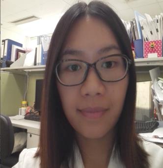
Li Ting is currently a PhD student in School of Chinese Medicine, Hong Kong Baptist University. She is working in exploring effective and safe traditional Chinese medicines or natural products against melanoma.
Abstract:
Constitutively activated signal transducer and activator of transcription 3 (STAT3) plays a critical role in melanoma development. A formula (SL) consisting of Sophorae Flos (SF) and Lonicerae Japonicae Flos (LJF) is documented as a remedy for subcutaneous ulcer, skin carbuncle and abscess. Plenty of active constituents in SF and LJF have been shown to inhibit STAT3 signaling and possess anti-melanoma property. This study aims to investigate whether SL exerts anti-melanoma activities by targeting STAT3 signaling. B16F10 melanoma allograft model was employed to examine the effects of SL on tumor growth. Human A375 and murine B16F10 melanoma cells were utilized to evaluate the effects of SL on cell proliferation, apoptosis, migration and invasion. A375 cells stably expressing STAT3C, a constitutively active variant of STAT3, were used to determine the involvement of STAT3 signaling in SL-afforded anti-melanoma effects. Oral administration of SL (1.2 g/kg) significantly inhibited tumor growth in B16F10 melanoma-bearing mice and inhibited phosphorylation of STAT3 and Src in tumor tissues. In melanoma cells, SL potently inhibited cell viability, induced apoptosis and suppressed migration and invasion. SL also remarkably suppressed activation of STAT3 and Src and STAT3 nuclear localization in melanoma cells. SL significantly lowered mRNA and protein levels of STAT3-regulated Mcl-1, Bcl-xL, MMP-2 and MMP-9. Overexpression of STAT3C in A375 cells diminished the effects of SL on cell proliferation, apoptosis and invasion. These results indicated that SL exerted potent anti-melanoma effects and these effects are partially due to the inhibition of STAT3 signaling.
Immaculate Mbongo Langmia
University of Malay, Malaysia
Title: Genetic consideration for treatment of skin cancer

Biography:
Immaculate M. Langmia is a research Fellow at the University of Malaya in Malaysia. She received a B.S. degree in Biochemistry from the University of Buea. She was awarded Msc. and Ph.D. in Pharmacogenomics from the University of Malaya. She has been active in the area of pharmacogenetic research and personalized medicine for over few years. Her current research involves pharmacogenomics of disease complications and biomarker discovery.
Abstract:
Skin cancer is the most common type of cancer in the world. Although the occurrence of skin cancer may vary from one geographical area to another, it is now clear that within the same environment, some individuals are more susceptible to skin cancer and likewise some patients respond quickly to anti-cancer therapy than others. According to genetic studies, these differences is due to genetic and epigenetics predisposing confounders. It is of importance for every government or institution to promote research that seek to provide solution for treatment and prevention of skin cancer in susceptible individuals. Understanding disease-associated genetic mutations, epigenetics changes and the biological pathways involved in the etiology of skin cancer may lead to improved treatment, novel therapeutics, and personalized medicine. This will help to reduce the number of deaths due to skin cancer worldwide.
- Cosmetology
Location: Melbourne, Australia
Session Introduction
Simaran Pal Singh Aneja
Aneja Skin Care Centre, India
Title: Efficacy of autologous platelet plasma combined with fractional ablative carbon dioxide laser therapy in the treatment of facial atrophic acne scars
Biography:
Dr. Simran Pal Singh Aneja has completed his MD Dermatology at the age of 28 years from Devaraj Urs University(Kolar, Karnataka) and MBBS from Sri Ramachandra University(Chennai). He is the director of Aneja Skin & Hair Centre, a premier Skin & Hair Centre.
Abstract:
Ablative Fractional Co2 Laser therapy is considered by most as gold standard for atrophic acne scars although it is associated with prolonged erythema at treated site, longer downtime which may affect the daily lives of patients. Autologous Platelet-rich plasma (PRP) is known to enhance wound healing and has been shown to act as a potential adjuvant with Fractional Co2 laser for the correction of acne scars. The objective of this study was to evaluate the synergistic effect of autologous platelet-rich plasma (PRP) combined with fractional CO2 laser therapy for acne scars. 55 patients with moderate to severe atrophic facial acne scars of different morphologies were enrolled . 48 of them completed the study. Of those who completed, each underwent 3 sessions of PRP combined with fractional co2 laser therapy at 6-8week intervals. Clinical photographs were taken and patients were assessed at each session. Final assessment was done 5 months after the last session using a four-point scale for clinical improvement of skin smoothness .More than 75% improvement was labeled as excellent response , 51-75% as good, 26-50% and 0-25% as fair and poor respectively . 12 patients (25%) showed excellent improvement 22(45.8%) and 14(29.2%) patients demonstrated good and fair response respectively. Average duration of operated site erythema and edema was 3-5 days. Two patients developed post inflammatory hyperpigmentation which lasted 6-12 weeks. PRP with ablative fractional co2 laser therapy is a good combination for treating acne scars with higher patient satisfaction for clinical improvement as well as shorter downtime .
- Dermatological Diseases and Disorders
Location: Melbourne, Australia
Session Introduction
Zainab Sawafta
Queen Elizabeth Hospital, UK
Title: Specialist excision of basal cell carcinoma leads to better outcomes

Biography:
Zainab Sawafta has completed her MBBS from the University of York and is currently in her second year of Medical training. She has completed her first year of training in Queen Elizabeth Hospital Birmingham, which is recognized as one of the leading hospitals in Europe. She is currently continuing her training in Birmingham. She is very enthusiastic about pursuing a career in dermatology in the near future and has carried out various projects and voluntary placements in the field.
Abstract:
Introduction: Basal Cell Carcinoma (BCC) is responsible for a significant healthcare burden, particularly among Caucasian populations. Its locally invasive and destructive potential results in high morbidity, if left untreated. The gold standard for this is surgical excision with histological analysis of margins. In the UK, BCC excisions are carried out in both primary and secondary care settings. Success of therapy is assessed through evaluation of excision margins, which we have compared between specialties.
Methods: A retrospective analysis of BCC histology reports was carried out over four months, analyzing excision margin based on specialty and operator experience. Biopsies were excluded. Data was taken from a District General Hospital histopathology laboratory, which analyses BCC specimens from Dermatology, Plastic Surgery, Oculoplastics, ENT and GP Departments.
Results: 549 patients had 629 lesions excised. Excisions carried out by specialty were as follows: 516 by Plastic Surgery, 78 by Dermatology, 26 by General Practice, 6 by Oculoplastics and 3 by ENT. Dermatology completely excised 86% of lesions with 8% less than 1 mm margin and 6% involving margins. Similarly, 85% of lesions were completely excised by Plastic Surgery, 10% less than 1 mm and 4% involving margins. GP completely excised 63% with 27% less than 1 mm and 8% involving margins. 18% of incomplete excisions were carried out by consultants.
Discussion: Findings show that specialties with higher caseload, therefore greater operator experience exhibit more favorable results when it comes to BCC excisions, compared to primary care. Consultants were far less likely to have incomplete excisions, compared to trainees or GPs.
Salim Musa Mulla
Mulla Ayurvigyan Hospital, India
Title: V-AT-A Vitiligo - Ayurvedic Treatment Approach

Biography:
Dr. Salim Mulla has completed his Ayurvedacharya – BAMS from Shivaji University , Kolhapur in 1998. He completed M.S.(Ayurvedavachaspati) in 2004 from University Of Pune. He has done studies in Medical Genetics from Maharashtra University of Health Sciences in 2008. Also done studies in Preventive Ayurvedic Cardiology & Panchakarma from Mahatma Gandhi University Meghalaya & Madhavbaug Institute Thane in 2013. Running Speciality Ayurveda Hospital at Islampur - Sangli Maharashtra. He has published 3 papers in reputed journals. He is engaged in research work in obstetrics & gynaecology, infertility, vitiligo & Ayurveda. He has presented scientific Papers at various National & International seminars & conferences.
Abstract:
This is a single blind randomized clinical study on patients of vitiligo , a disease rather difficult for cure. Aetiology is multifactorial, it may be hereditary , autoimmunue, hormonal imbalance , dietary, stress , secondary to other systemic diseases like diabetes mellitus, hypothyroidism etc. This study is carried out on 200 outdoor patients suffering with vitiligo. Patients from both sexes, from age group five to seventy years complaining mainly as white patches are studied. Patients are treated with ayurvedic polyherbal powder mixture containing Psoralia corylifolia ( Bakuchi) as main ingredient supplemented with local application and phototherapy (natural). Most of the patients undergo intermittent bodypurification i.e. shodhana – panchakarma treatments like vamana ( therapetic emesis ) , Virechan ( purgation), Basti ( enematous treatment for Vaatadoosha) , raktamokshana etc as per ayurvedic guidelines. Duration of treatment is of one month to one year depending upon response & requirement for the treatment. Followup is biweekly to monthly. The response to treatment is observed in terms of reduction in area of depigmentation after treatment. Complete cure is noted in 12 % patients. Good result is noted in 18 % cases , moderate in 45 % cases and mild result in 25% cases noted. The result is statistically highly significant at 0.1% level. No major side effects of the treatment given observed. The treatment is effective in all types of vitiligo. The results of internal medications & local treatment are aggrevated specially after panchakarma treatment.
Friend Philemon Liwanag
Southern Philippines Medical Center, Philippines
Title: De novo histoid leprosy manifesting over a tattoo site
Time : 14:45-15:15
Biography:
Friend Philemon M Liwanag has completed his Doctor of Medicine degree at the Manila Central University-FDT Medical Foundation. He is a currently a second year Dermatology Resident at the Southern Philippines Medical Center in Davao City, Philippines.
Abstract:
Histoid leprosy is recognized as a rare form of leprosy with a heavy bacillary index. De novo is a peculiar type of histoid leprosy characterized as an initial clinical presentation without previous treatment with multidrug therapy. Further, published reports have shown the possible participation of skin other than respiratory droplets in leprosy transmission. We report a case of de novo histoid leprosy in a 30 year old man, who initially presented with a hyperpigmented papule which progressed into a shiny nodule over a tattoo site on his left upper arm. The lesions became generalized with symmetrical distribution. Histopathological studies revealed findings consistent with the diagnosis. Histoid leprosy was first described in the Philippines, which exemplifies the challenge to leprosy eradication. Thus, this finding that tattooing as likely cause of inoculation of histoid leprosy in this post-elimination era generates important consideration.
- Cosmetic Dermatology
Location: Melbourne, Australia

Chair
Simaran Pal Singh Aneja
Aneja Skin Care Centre, India
Session Introduction
Simaran Pal Singh Aneja
Aneja Skin Care Centre, India
Title: Efficacy of autologous platelet plasma combined with fractional ablative carbon dioxide laser therapy in the treatment of facial atrophic acne scars
Biography:
Simran Pal Singh Aneja has completed hid MD Dermatology at the age of 28 years from Devaraj Urs University(Kolar, Karnataka) and MBBS from Sri Ramachandra University (Chennai) . He is the director of Aneja Skin & Hair Centre, a premier Skin & Hair Centre.
Abstract:
Ablative Fractional Co2 Laser therapy is considered by most as gold standard for atrophic acne scars although it is associated with prolonged erythema at treated site, longer downtime which may affect the daily lives of patients. Autologous Platelet-rich plasma (PRP) is known to enhance wound healing and has been shown to act as a potential adjuvant with Fractional Co2 laser for the correction of acne scars. The objective of this study was to evaluate the synergistic effect of autologous platelet-rich plasma (PRP) combined with fractional CO2 laser therapy for acne scars. 55 patients with moderate to severe atrophic facial acne scars of different morphologies were enrolled . 48 of them completed the study. Of those who completed, each underwent 3 sessions of PRP combined with fractional co2 laser therapy at 6-8week intervals. Clinical photographs were taken and patients were assessed at each session. Final assessment was done 5 months after the last session using a four-point scale for clinical improvement of skin smoothness .More than 75% improvement was labeled as excellent response , 51-75% as good, 26-50% and 0-25% as fair and poor respectively . 12 patients (25%) showed excellent improvement 22(45.8%) and 14(29.2%) patients demonstrated good and fair response respectively. Average duration of operated site erythema and edema was 3-5 days. Two patients developed post inflammatory hyperpigmentation which lasted 6-12 weeks. PRP with ablative fractional co2 laser therapy is a good combination for treating acne scars with higher patient satisfaction for clinical improvement as well as shorter downtime .

